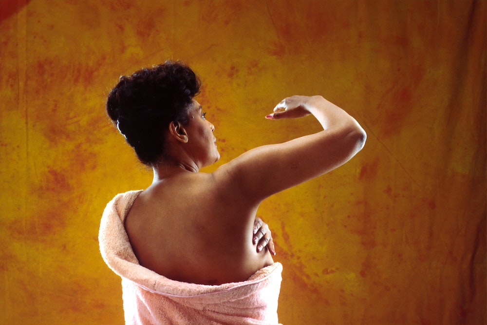
Woman holds breast cancer pink ribbon, pointing at it to encourage others to understand mammogram screening guidelines.

A Guide to Mammograms: What Age Do Mammograms Start, What Are Mammograms Like, and More

Firstly, we want to congratulate you on doing the necessary research to be ready for your mammogram! Yes, getting a mammogram for the first time is intimidating, but if you’ve successfully scheduled and overcome a pap smear, you are capable of anything.

Mammograms are used to detect early signs of breast cancer. Breast cancer is most treatable in its early stages, which is why preventative checks are so crucial for women. Mammograms are similar to getting an X-ray of your breast tissue. They allow doctors to physically see inside breasts to check out what, exactly, is happening in there. They’re especially useful because they really give the doctor everything they need to know - these days, mammograms capture even the most minute details, so they’re highly effective. Trust us, scheduling a mammogram is worth every woman’s time.

So, what’s the difference between a mammogram and an ultrasound?

An ultrasound uses high-frequency sound waves that bounce off the tissues within our bodies, which a computer then converts to an image.

A mammogram, on the other hand, is a low-dose version of an X-ray, which is why metal plates are needed during the procedure. The electrodes released during the mammogram have to bounce off the surrounding metal to capture the image of the mammogram, showing healthcare providers any breast cancer or other breast diseases present. Sometimes, both mammograms and ultrasounds are used in tandem to help the doctor understand what is going on within the breast.

Ultrasounds are not a replacement for mammograms of breast cancer, but aid doctors in checking any abnormal areas found in patients with mammograms of dense breast tissue. They are used in addition to a mammogram, but never replace one entirely!

Nowadays, doctors will decide to administer one of three types of mammograms: film-screen mammography, digital mammography in 2D, and digital mammography in 3D.

What is digital mammography in 2D?

This practice takes two images of each breast and uses them to create a 2D image of each. It was standard practice until 2011.

What is digital mammography in 3D?

Also known as digital breast tomosynthesis, digital tomosynthesis, or just tomosynthesis, 3D digital mammography uses several images of each breast to create a detailed 3D image of the breast. This has become standard care for breast cancer after gaining approval from the FDA in 2011. Studies show that 3D digital mammography finds more cancers than the traditional 2D method and gives fewer false positives.

What is film-screen mammography?

This type of mammogram produces black-and-white images on film, takes a little more time to create, and was more commonly administered in the past.

Doctor listens to woman's heart with a stethoscope before her first mammogram.

What’s the age of the first mammogram in the U.S.?

The United States Preventative Services Taskforce recommends that women begin to regularly schedule screening mammograms at the age of 40. After, mammograms should be scheduled every one to two years for a breast check-up if there are no symptoms found.

When should I begin self-examining my breasts?

This is another crucial preventative step! And it’s a quick and simple process. Young women should begin self-examining their breasts as soon as they mature and their breasts stop growing, or around the age of 20. Read through this helpful guide on how to self-examine your breasts.

When women examine their breasts regularly beginning at a young age, they are more likely to discover any irregularities or changes before they develop into something more serious. Self-examination is another fantastic preventative practice all young women should include in their weekly schedules.

Waiting until the week after your period to conduct self-examinations is recommended to avoid any pain or abnormalities caused by hormone fluctuations and swelling.

When self-examining breasts, always look out for…

  • Lumps that are new, growing, or changing shape.
  • Swelling or thickening of a certain area or part of the breast or armpit.
  • Nipple abnormalities like reddening, flaking, pulling in, or discharge.
  • Pain that is sudden, irregular, or creates constant discomfort.
  • Changes in size that are sudden or seem out-of-the-blue, unrelated to hormonal fluctuations or periods.

If you discover any of these symptoms of breast cancer, reach out to your doctor or schedule a diagnostic mammogram with a doctor near you to check your breasts as soon as possible.

Take a closer look at mammogram screening guidelines.

So, whether it’s your first time getting a mammogram at age 40 or taking preventative steps and scheduling a diagnostic mammogram early,…

Doctor aids woman to get into correct position for her mammogram.Here’s Our Step-by-Step Guide to Your First Mammogram

Schedule a Mammogram Appointment:

  • Ask your doctor or do some research to find a reputable mammography facility or clinic in your area.
  • Call or register online to schedule an appointment.
  • Make sure they specialize in mammograms and have qualified radiologists!

Timing:

  • Avoid scheduling the appointment during the week before your period when your breasts may be more sensitive. Although mammograms are not invasive, pressure is applied to the breast which can create discomfort.

Clothing and Comfort:

  • We recommend wearing a two-piece outfit, as you'll need to undress from the waist up.
  • Avoid wearing deodorant, lotions, or powders on your upper body, as they can interfere with the imaging.

Communicate and Share:

  • Sharing your medical history, prior surgeries, or family history of breast cancer can help doctors provide the best care.
  • Always inform the technician if you have breast implants or have had any breast-related procedures or concerns.

How long do mammograms take?

  • Roughly 20 minutes!
  • You’ll be asked to undress from the waist up and will be given a gown to wear during the procedure.

The Procedure:

  • First, the technician will position your breast between two metal plates on the mammography machine.
  • Then, the plates will compress your breast for a few seconds to get the clearest images. While it may feel uncomfortable or slightly painful, it shouldn't be overly painful. Communicate any discomfort you feel to the technician.
  • Because mammograms usually involve two images of each breast taken from different angles, the technician may reposition your breast between images.

After the Procedure:

  • Once the images are taken, you'll be asked to wait while the technician ensures the images they captured are high-quality.
  • Once the quality of the images is confirmed, you can get dressed.

             Some women may experience mild bruising or temporary discomfort. This should subside quickly.

How long does it take to get mammogram test results?

  • You’ll receive your results within a few weeks after your radiologist interprets your mammogram and sends the results to your healthcare provider.

           Don't panic if you're called back for additional tests, as most callbacks do not indicate breast cancer.

What to Do Before and After a Mammogram:

  • Stay relaxed and take deep breaths before the procedure - always communicate discomfort and pain to your doctor!
  • Follow all instructions given to you by the mammography facility.
  • Afterward, continue to schedule regular mammogram screenings as recommended by your healthcare provider.
  • Know your mammogram schedule and never skip routine screenings.

What Not to Do Before and After a Mammogram:

  • Don't wear metallic accessories or clothing items, as they interfere with the mammogram images.
  • Try not to schedule your mammogram at a time when your breasts are likely to be sore or swollen to avoid discomfort.
  • Don't be afraid to ask questions and voice any concerns to your technician or radiologist.

We hope this guide on how to prepare for your first mammogram helps; that’s what we’re here for! At Pawsitively 4 Pink, our passion is providing direct financial support for local, underserved women fighting breast cancer. Please contact us to find out more about ways to help, or donate directly.


I Found a Lump in My Breast...Now What?

You’re in front of your dresser and your hand runs through your chest. You feel something and just to be certain, you touch your breast again and this time, you’re sure that you felt a lump. Most women self-examine their breasts and once they find a lump, they freeze. Now the question is, what do you do when this happens?

First and foremost, don’t panic. Research shows that 85%  of breast lumps are benign, which means they are noncancerous, especially in women who haven’t yet hit 40. Women over 40 are normally encouraged to have regular mammograms and if those mammograms always turn negative, the odds are even higher that the lump you felt isn’t cancerous.

According to most obstetricians, before a palpable lump appears on a woman’s breast, something will already have been detected during a mammogram screening. Despite most women knowing this, the worrying and stress it has not stopped anyone from worrying once they feel a lump. The only way to be sure is to differentiate between a benign lump and a breast cancer lump.

Understanding your breasts

Your breasts consist of fat, blood vessels, nerves, glandular tissue, fibrous connective tissue, ducts, and milk-producing lobules. All these make your breasts quite lumpy. The major difference between a benign lump and a cancerous lump is that a benign one is fluid-filled and rolls between your fingers. A cancerous lump is normally hard and feels stuck in one place.

The other difference has to do with pain. Breast cancer, in most cases, doesn’t cause pain. Inflammatory breast cancer is an exception to this, as it comes with symptoms of aching or burning in the breast. Benign conditions might also be accompanied by painful symptoms, but at times, this isn’t the case.

The only sure way to know the nature of a breast lump is via medical tests such as a mammogram, fine needle aspiration, or ultrasound. These tests should be done at a medical facility that has all the equipment and with experts who can read the results.

Understanding the tests

What is a mammogram?

An X-ray examination of the breast is referred to as a mammogram. It is a process used to detect breast diseases or any breast problems, such as nipple discharge, lumps, or pain. This procedure enables the detection of benign tumors, cysts, and breast cancers before they can be detected via touch. All women are encouraged to frequently do a mammogram, whether or not they have breast health-related issues.

There are factors or conditions that may affect your mammogram results, such as

  • Having had a previous breast surgery
  • Hormonal breast changes
  • Deodorant, lotions, creams, or talcum powder applied on breasts or under the arms
  • Breast implants; these prevent the complete visualization of the breast area, hence hiding tissue

You should inform your medical examiner if any of these apply to you so that they can make the required preparations.

How to prepare yourself for a mammogram

The first thing is scheduling a date for the mammogram. It is advisable to do this two weeks before your period starts because breasts do become tender a week before and during menstruation. Once again, if you have breast implants, please notify the facility when scheduling the exam.

Expectant mothers or those who think they might be pregnant should consult a medical doctor before scheduling a mammogram. Discuss all options with your doctor. If you are breastfeeding, you should mention this to the mammography technician.

Avoid using lotion, perfume, deodorant, or powder on the day of the examination. Note that you'll have to remove clothes from the waist up, so wear something that you can easily get out of. It is also advisable to leave all jewelry and valuables at home. The health care provider or technologist will request other specific preparation based on your medical condition.

What is a fine-needle aspiration or FNA?

There are situations where a mammogram can’t prove that a certain body area is cancerous even though there are high chances that it is. In such situations, the tissue will be extracted via a needle for biopsy. The tissue will then be scrutinized under a microscope to determine if it is cancer or not.

FNA is just one of the several breast biopsy procedures as the type of biopsy done depends on the size and location of the breast lump.

How to prepare yourself for a biopsy

The entire procedure should be explained to you by the technologist and you should ask all the questions you might have. A consent form will be provided, but you should go through it carefully before signing anything. Once again, seek clarification on points that you don’t understand.

If your breast is numbed using local anesthesia and you are awake, then you don’t have much preparation. If general anesthesia will be used to put you into a deep sleep, then you will have to fast before the surgery. Your surgeon will explain everything before the procedure.

Once again, apply nothing to your breast or underarms. Inform your healthcare provider if you suspect you could be pregnant or you are pregnant. You should also inform them of any allergies. Perhaps you could be allergic to latex or anesthesia. You should also let them know if you are on any medication or taking supplements. This is also the time to say if you have a history of bleeding disorders.

Since you will be drowsy and groggy after, ensure that someone is on standby to drive you home.

What is an ultrasound?

When sound waves are used to examine your breasts, we refer to the method as a breast ultrasound. It will help the medical examiner spot any issues and also see how well blood flows to your breast areas. Ultrasounds are normally used when there is a change that fails to show up in a mammogram.

A breast ultrasound is not normally used to detect breast cancer, the reason being that it may fail to detect some early signs. For example, one early cancer sign it might not detect is micro-calcifications, which are small calcium deposits. So in what cases are an ultrasound method used?

  • When the breast tissue is really dense and the mammogram cannot see through it
  • When pregnant. Ultrasound doesn’t make use of radiation like mammography.
  • You are below 25 years of age
  • When there is a need to look at the surrounding lymph nodes. This is essential when removing cyst fluid or guiding a needle during a biopsy.
  • The health provider may recommend other reasons

How to prepare yourself for an ultrasound

Your healthcare service provider should explain the entire procedure to you. Don’t be afraid to ask all the questions that you may have. There are situations where you might be given a consent form to sign before the test. It is important that you go through the form carefully and ask for clarification when something is not clear.

There is no need to stop eating or drinking before the test. Just relax and please, don’t take any medicine for it. Breathe easy. Once again, avoid putting lotion, deodorant, or any other substances on your breasts that day. You should also wear clothes you can easily take off and that allow the technologist to easily examine your chest area. Some gel will be applied to your skin during the test. Follow any other instructions as required during the test.

Information is power, so when you feel a lump on your breast, seek medical advice as ll breast lumps should be evaluated by a medical professional. Once the cause of the lump is established, treatment can begin. So, finding a lump on your breast is not always a cause for alarm, it is a cause for taking the right action.

If you’d like to help make a difference in the lives of women experiencing breast cancer, you volunteer with us a Pawsitively 4 Pink or make a small donation.